


St. Mary Medical Center in Middletown is now offering a 3D ultrasound for breast cancer screenings.
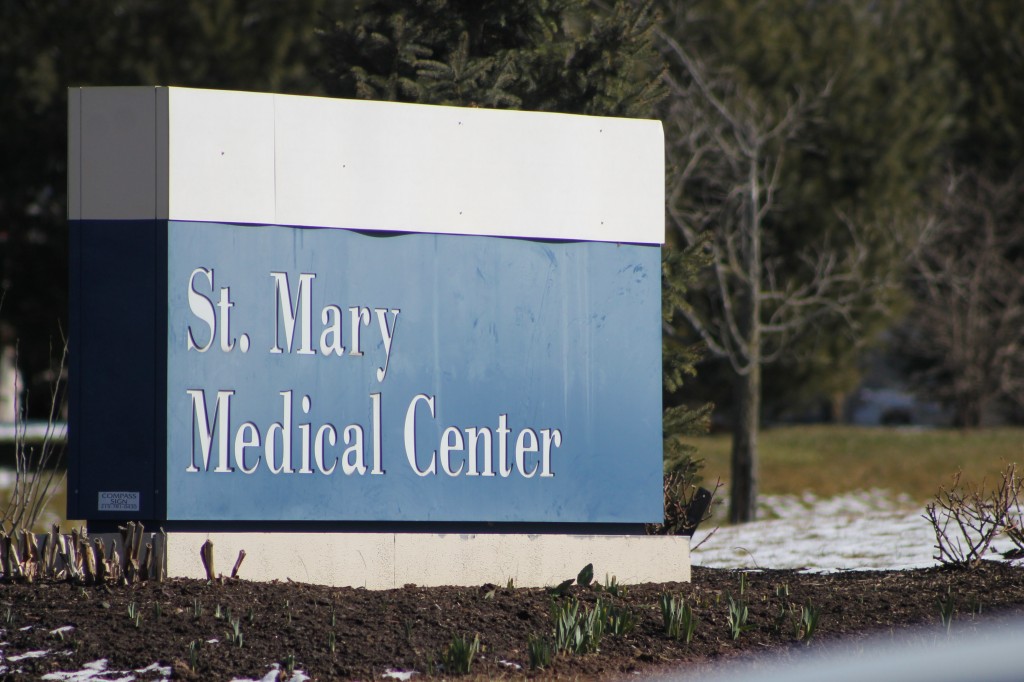
Credit: Tom Sofield/LevittownNow.com
The hospital’s breast center is using advanced digital mammography coupled with computer-aided detection (CAD) and produce excellent quality images, according to a press release. The dual method system helps doctors see into dense breast tissue that can sometimes make it difficult to detect some cancer lesions.
Advertisements
“Women who have dense breasts or other risk factors for cancer potentially benefit the most from additional screenings, such as 3D ultrasound which provides critical details for a precise diagnosis without incurring additional radiation to the patient, ” Dr. Gustavo Sanchez, chief of St. Mary imaging, said in a statement. “This new technology provides a more realistic view of the anatomy of the entire breast.”
Here’s how the hospital explains the 3D breast ultrasound:
The 3D Total Breast Ultrasound provides a slice-by-slice evaluation of the breast anatomy, from the skin to the thoracic wall, a view not available using conventional 2D ultrasound. Automated Breast Volume Scanning (ABSV) uses computerized pathways to scan every part of the breast and reconstructs the images for evaluation by a dedicated mammography radiologist. An entire 3D ultrasound of both breasts is comfortable, painless, and takes about 20 to 30 minutes to perform.
Breast density is the ratio of fat to fibroglandular tissue in the breast. Dense breast tissue is a common finding and is not abnormal. But for those women who may have dense breast tissue on mammography, a mammography alone has been shown to have limited effectiveness in visualizing breast cancer. In compliance with the Breast Density Notifications Act enacted in Pennsylvania in February 2014, all patients who have a mammogram performed at St. Mary now are informed about their breast density in the results letter they receive from the Breast Center.
Women who have both dense breast tissue and other risk factors may be at increased risk for breast cancer. A 3D ultrasound evaluation provides greater detail of the breast anatomy that can aid in earlier detection and a more accurate diagnosis. Women who have been notified that they have dense breast tissue are encouraged to consult with their physician about the benefits of additional imaging after a mammogram.
For more information, call the breast center at 215-710-5393.

